Context Notes: By combining virtual reality with a technique called expansion microscopy, scientists are enlarging, exploring and analyzing cell ... Unprecedented views of the interior of cells and other nanoscale structures are now possible thanks to innovations in expansion ...
Proexm For Tissues Gelation Demonstration - Guide Important Details
This browsing page explains Proexm For Tissues Gelation Demonstration through quick context, useful references, alternate wording, and broader search ideas so readers can continue into related pages with clearer context.
In addition, this page also connects Proexm For Tissues Gelation Demonstration with for broader topic coverage.
Guide Important Details
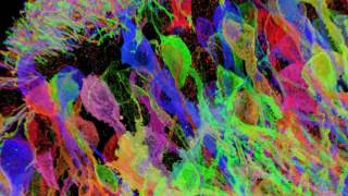
By combining virtual reality with a technique called expansion microscopy, scientists are enlarging, exploring and analyzing cell ... Unprecedented views of the interior of cells and other nanoscale structures are now possible thanks to innovations in expansion ... Brainbow-expressing mouse hippocampal brain circuitry, processed via the
Guide Summary
Brainbow-expressing mouse hippocampal brain circuitry, processed via the This series of How-To videos walks you through best practices for preparing
Helpful Background for Readers
This part keeps Proexm For Tissues Gelation Demonstration connected to practical references instead of leaving it as a single isolated phrase.
Helpful Reminders for Readers
Before relying on any single result, compare related pages and verify important facts from stronger sources.
Important details found
- This series of How-To videos walks you through best practices for preparing
- By combining virtual reality with a technique called expansion microscopy, scientists are enlarging, exploring and analyzing cell ...
- Unprecedented views of the interior of cells and other nanoscale structures are now possible thanks to innovations in expansion ...
- Brainbow-expressing mouse hippocampal brain circuitry, processed via the
How readers can use this page
Readers use this page when they need a simple summary for Proexm For Tissues Gelation Demonstration before checking official or primary sources.
Common Questions
What details can change around Proexm For Tissues Gelation Demonstration?
Dates, prices, policies, availability, providers, software versions, and public details may change over time.
What supporting details help explain Proexm For Tissues Gelation Demonstration?
Comparison helps readers avoid narrow results and find the angle that best matches their intent.
How should readers use this page?
Use this page as a starting point, then open related entries or official sources when exact details matter.
What makes Proexm For Tissues Gelation Demonstration easier to understand?
Clear headings, short explanations, practical notes, and related entries make Proexm For Tissues Gelation Demonstration easier to scan and compare.